What is a spider?
 Click to enlarge image
© ©Australian Museum
Spiders have:
- two main body parts, the head and thorax combined called the cephalothorax and an abdomen
- eight walking legs
- simple eyes; spiders usually have eight eyes (some have six or fewer), but few have good eyesight.
- jaws adapted for tearing or piercing prey
- a pair of pedipalps
- abdominal silk spinning organs
- anterior abdominal genital opening.

© Australian Museum
© Australian Museum
Spider body parts
A spider's body is in two sections. The head and thorax, bearing the eyes, mouthparts and legs, are fused together to form the cephalothorax. This is joined by a slim waist (pedicel) to the second body section, the abdomen, on which are found the silk spinning organs (spinnerets), the reproductive openings and the breathing organs (book lungs and/or tracheae).
The cephalothorax
The cephalothorax is covered above by a hard cuticular plate called the carapace - much like the hard 'shell' covering a crab.
On the outside of the cephalothorax are
- the simple eyes - usually eight (sometimes six), are commonly arranged in two rows along the front of the carapace (although eye arrangement and sizes vary).
- the fovea - a depression in the middle of the carapace which is the internal attachment point for thoracic muscles.
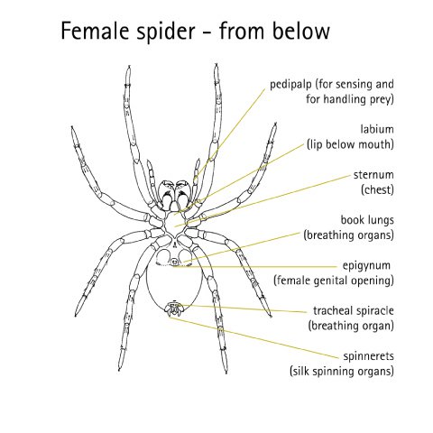
- the mouthparts - two large jaws with their piercing fangs (the chelicerae), while behind the jaws on the underside there are two small cuticular plates (flattish blocks of cuticle) - an upper plate, the labrum (upper lip) which is hidden by a lower plate the labium (lower lip), clearly visible from below behind the jaws. These two plates form the roof and floor respectively of the tube-shaped mouth which opens just behind the jaws. A pair of plate-like maxillae flank the labium - each has a food cutting row or patch of teeth on the front end.
- the pedipalps - that help with food handling, touch and taste sensing and, in male spiders, are modified as mating organs.
- the legs - four pairs of jointed legs with two to three terminal claws. The two-clawed spiders are hunters (e.g., jumping spiders, huntsman spiders, ground spiders), most with thick hair brushes (scopulae or claw tufts) on the ends of the legs - these improve traction on smooth or sloping surfaces like leaves or tree trunks. Many of the three-clawed spiders are web builders, often with claws and hairs modified for silk handling (e.g., orb-weavers, gum-footed web spiders, lace-web spiders).
On the inside of the cephalothorax are
- the muscles - to help move the jaws and limbs. Muscles from limb, gut and carapace attachments are all connected to the central endosternite, an internal non-chitinous skeletal plate.
- the brain ganglia - a mass of nerve tissue.
- the venom glands - to produce venom to kill prey
- the muscular stomach - to pump the liquid food up into the oesophagus (food pipe) and pharynx (throat) and move it along the gut. The end of the fore-gut forms the stomach. Diverticula (outgrowths) that extend into the legs are also in the cephalothorax.
The abdomen
The abdomen is usually covered with a thinner or more flexible cuticle - this allows for expansion with feeding or when eggs are developing. The thin waist or pedicel separating it from the cephalothorax allows movement of the abdomen, for example, during silk spinning and mating displays.
On the outside of the abdomen are
- the book lung covers - to protect the delicate organs inside
- the gonopore or genital opening - from which eggs or sperm are released is placed in the genital groove between the front pair of book lungs. In most female araneomorph spiders there is another separate, plaque-like mating opening, the epigynum.
- the spinnerets (silk spinning organs) - usually four or six in number, and the terminal anal tubercle on which the gut ends at the anus.
On the inside of the abdomen are
- the book lungs - the breathing organs. Small openings called spiracles lead into air filled cavities into which the thin, leaf-like lamellae of the book lungs project - like rows of book pages. The outer surfaces over which air passes are covered by a very thin cuticle from which peg-like struts project, keeping the lamellae from collapsing. Blood (haemolymph) circulates within the lamellae and gaseous exchange between blood and air occurs across their thin walls. There are two pairs of book lungs in mygalomorph (and some araneomorph) spiders. Most araneomorphs have the front pair of book lungs only, the rear pair being replaced by fine, cuticular tracheal tubes that divide within the body and allow more efficient gas exchange. A few tiny spiders living in moist, sheltered habitats have no breathing organs, gas exchange taking place directly across the thin body cuticle.
- silk glands - to produce the liquid protein that makes the silk
- reproductive organs (ovary or testes).
- the heart - lies in the midline of the body, where it can be seen beating through the dorsal cuticle. The blood circulation is open - that is, the blood vessels from the heart open into the body space, bathing the tissues and organs in blood which then gradually circulates back to the heart.
- the hind-gut and its diverticula - where absorption of nutirents occurs into tissues. The hind-gut has a sac into which excretory organs called malphigian tubules (the spider's 'kidneys') open.
Jaws and fangs
In mygalomorph spiders (trapdoor and funnel-web spiders) the large jaw bases project forward in parallel with their fangs folded back side-by-side underneath. To bite their prey these spiders must raise the front of the body, allowing the fangs to open like a pair of daggers for a downward strike. In the more common araneomorph spiders (redbacks, wolf spiders, etc.) the jaws are slung vertically under the front of the carapace. The fangs are hinged laterally and bite cross-ways against each other, like pincers. This is a more efficient arrangement for seizing and manipulating prey, especially on a web.

© Australian Museum
Spider Skin
Like other arthropods, the spider's body is covered with a more or less rigid 'skin' or cuticle (the exoskeleton) made of protein and chitin. The spider cuticle consists of several layers, the outermost being toughest, covered with a thin surface wax layer that helps reduce water loss from the body. The cuticle provides internal attachment points for the muscles and helps in the regulation of blood pressure. While it is hard and protective externally, the cuticle must still accommodate the spider's sense organs - in the form of various types of innervated (supplied with nerves) hairs and pits, as well as the eyes. The cuticle even extends internally, lining the fore gut (mouth to stomach) and hind gut, the tracheal (breathing) tubes and the female's sperm storage organs (spermathecae).
To allow the spider to grow the entire cuticle must be shed periodically, a process known as moulting. A new larger cuticle is first made underneath the old one, the old one splits and the spider climbs out. The new cuticle is very soft and most spiders will not move untill the cuticle hardens.
Spider skeleton
A spiders exoskeleton encloses the blood-filled body space. Confined within this semi-rigid space, the blood pressure can be varied by changes in heartbeat frequency or contraction and relaxation of muscles, notably the strong thoracic muscles. Together, the cuticle and the blood make up a pressurised unit known as the hydrostatic skeleton. This is important in maintaining body shape (turgor) and function.
Life and death
The ability to vary blood pressure is important in functions as diverse as moulting and movement. During moulting, increased heart rate results in blood pressure increases that help split open the weakened cuticle. Limb extension during movement is achieved mainly through the contraction of strong thoracic muscles which increases thoracic blood pressure and causes the limbs to extend outwards. This explains why spiders have many flexor muscles for bending their limbs inwards, but fewer extensor muscles for stretching them outwards - they're just not needed as much. It also explains why injured or dead spiders always have their legs bent inwards - they can no longer control their blood pressure and this allows the strong flexing muscles to dominate and pull the legs in under the body. This 'death position' is imitated by spiders that escape predators by dropping from a web to the ground - only this time the legs are deliberately flexed in while playing dead.
